Ovine Progressive Pneumonia
Ovine progressive pneumonia (OPP) is a slowly progressing viral disease of sheep. The disease can affect many organ systems including lungs, joints, udder, and central nervous system. Because it is a disease where there are no immediate clinical signs upon infection, the economical impact on sheep producers may be hard to assess especially if the incidence of clinical disease is low. The respiratory form of OPP was first described in the United States in 1923 by Marsh in Montana, and called "progressive pneumonia." However, a 1915 report in South Africa described a similar disease called "Graaff-Rinet" disease. It was later found that this disease condition was identical to the disease described by Marsh. In Iceland in 1947, a pulmonary form of the disease was identified. The disease was called "maedi" meaning dyspnea or difficult breathing. Later in 1957, Iceland identified yet another form of the disease that caused paralysis. This form was called "visna" which means wasting. This resulted in the name "maedi-visna" virus, which is used in many countries to describe the same disease syndrome as OPP.
Virus Characteristics
OPP is caused by a lentivirus (lenti = slow), which is a member of a group of viruses called retroviruses (retro = reverse). This group of viruses; retroviruses, share a common characteristic in that they all utilize the hosts genetic machinery to replicate by incorporating their own reverse transcriptase enzyme. The retrovirus family of viruses cause slow progressive diseases that affect many species of animals and humans (AIDS). However, with some exceptions, most of these viruses are species specific (they only infect one or two closely related species). Experimentally, OPP has shown to infect goats. Lentiviruses can induce disease in many different organ systems and cause life long infections that may or may not cause death to the host. They do this by infecting and interfering with the host's immune system. This prevents the host immune system from clearing the infection, and makes it extremely difficult for scientist to develop vaccines for these viruses. Although studies show conflicting results, some breeds of sheep appear to be more susceptible than others to developing clinical signs to OPP virus, and some strains of the virus probably cause a more severe form of the disease. Sheep of the Texel, Border Leichester, and Finnish Landrace breeds appear to develop more frequent and more severe forms of the disease than do Columbia, Rambouillet, and Suffolk breeds. All breeds of sheep are susceptible to OPP viral infection.
Prevalence
With the exceptions of Australia and New Zealand OPP is present in all major sheep producing countries. In North America, infection is common in all breeds, sexes, and ages of sheep. Antibodies to OPP are not protective against the disease; therefore if an animal has antibodies to OPP after six months of age then they are considered infected with the OPP virus and are carriers of the virus. In the Western United States, antibodies to the virus are more frequently found than in other parts of the United States. This could be due to the fact that there are more sheep in the Western United States than in other areas of the U.S.
Clinical Signs of Disease and Associated Lesions
In the early stages of infection no clinical signs of disease are apparent. This is because of the nature of the virus as discussed above. In fact, most animals do not exhibit clinical signs of disease. However, those animals that do exhibit clinical signs usually are at least two or more years of age. The clinical signs can manifest in many different ways and can be attributed to involvement of one or more organ systems. Often the clinical signs are accentuated by times of stress such as lambing.
Early Stages of Infection
As stated before most animals do not exhibit signs of infection until two years of age. One of the most apparent clinical signs first noticed by producers is a general loss of body condition. This is often referred to as "thin ewe syndrome." The weight loss progresses in the face of a normal appetite. The exact pathophysiology of this ill thrift is not understood, but it should be understood that it is not limited to ewes.
Respiratory Tract Involvement
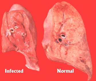
Involvement of the respiratory tract during the course of disease is common. The respiration rate at rest is increased, the animal tires easily, and is often found trailing the flock. These sheep are often referred to as "lungers." During the end stages of respiratory involvement, these animals will often become recumbent. Secondary bacterial pneumonia is a common sequela to the respiratory form of OPP, and is manifested by coughing and nasal discharge. At necropsy the lungs are consolidated: they have a rubbery consistency, do not collapse upon opening the chest, weigh 2-3 times normal weight, and have a dull grayish blue to grayish brown color. Histopathology shows that alveolar membranes where gas exchange takes place are thickened. The thickening is due to accumulations of lymphocytes and macrophages, with scar tissue and smooth muscle thickening also present. Smaller airways and blood vessels in the lungs are also infiltrated by lymphocytes. This can manifest grossly as nodules that are seen and felt within the lung tissue. Lymph nodes in the chest can be up to 10 times their normal size.
Udder Involvement
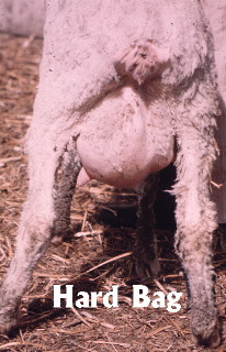
OPP viral infection can also infect the udder. This involvement becomes apparent as an enlarged firm udder that has reduced milk flow to no milk flow. This is often referred to as "hard bag." Milk production is decreased because of infiltration of lymphocytes and macrophages into the surrounding milk ducts where milk is produced. The changes found with udder involvement during the course of OPP virus infection are different from changes associated with mastitis. The udder is not hot or painful, nor are there abnormal milk secretions as are found in the case of mastitis. This is not to say that all cases of "hard bag" are caused by OPP. Other causes include plant estrogens, hormonal imbalance, and bacterial infection. The main difference is that the changes associated with udder involvement in OPP are generally irreversible, while the other causes may be reversible. There has been slight increases in milk production in OPP infected ewes a few days after lambing, but this transient slight increase is usually attributed to a slight decrease in udder swelling.
Arthritis
Arthritis; joint inflammation, has been associated with OPP virus infection. Swelling in one or more joints becomes apparent with the "knee" joint of the forelimb and the hock joints of the rear limbs most commonly affected. The arthritis is caused by a thickening and mineralization of the joint capsule and the subsequent bone and cartilage deterioration. The longer this inflammation persists the greater the damage to the joint and the greater the deterioration of the joint. Again, this arthritic change is preempted by infiltration of lymphocytes and macrophages into the joint.
Central Nervous System Involvement
Central nervous system involvement is the least frequent form of OPP seen in the United States. Early signs are muscle quivering and imbalance seen in the rear legs. This slowly progresses to total paralysis of the hind legs, which makes the sheep unable to stand. Histologically lymphocytes and macrophages are accumulated around blood vessels, and degeneration of nervous tissue is seen too.
Transmission
Since the virus is carried in macrophages, which are found in the tissue and fluids of the sheep, bodily secretions are sources of virus transmission. Secretions of the udder and lungs are thought to be the main source of virus transmission. Transmission from ewe to lamb via colostrum and milk has been documented. In addition, the longer the lamb stays with an infected ewe the greater the risk that the lamb will become infected. Infected ewes can also be the source of infection for lambs produced by uninfected ewes. In adults, the transmission of disease is thought to occur mainly through respiratory secretions, especially in confinement situations. Other bodily secretions such as blood, semen, and urine are not yet proven to transmit the disease, but should still be considered possibilities.
Diagnosis
Diagnosis of OPP can be difficult. Most sheep do not show any clinical signs, and those that do show clinical signs that are similar to other diseases. Therefore clinical signs alone are not often used to diagnose disease. Clinical signs are usually compared to serologic tests or necropsy findings to confirm the diagnosis of OPP. Since most producers would prefer that diagnosis be made ante-mortem, serologic testing is the most useful tool practitioners have to confirm a diagnosis, but it does have its limitations. The two most commonly used methods are agar gel immunodiffusion test (AGID), and enzyme linked immonosorbency assay (ELISA) test.
AGID is a less sensitive test and more prone to false negative results, but a positive AGID test
IS an OPP infected animal, whereas the ELISA test is more prone to false positive results. Both the AGID and ELISA tests perform at 96% accuracy. Nevertheless, because of the greater ease in performing this AGID test compared to ELISA it is usually the test of choice for screening flocks. Detection of OPP virus can also be done by culturing, but this is an expensive procedure and may require up to 12 weeks before results are available.
Treatment and Control
Treatment of OPP has not been found very effective. Because most sheep die due to secondary bacterial pneumonia, antibiotics can be used to prolong the sheep's life a few weeks or months. But, one should realize that these animals are still shedding the virus and possibly infecting the rest of the flock. Control of the disease can range from removing lambs from the ewe at birth and feeding them pasteurized colostrum and milk replacer to periodic testing and culling of positive animals. Using these methods some producers have been successful in eradication of the disease. Possibly, the most important aspect in controlling the spread of disease is to get replacement animals from flocks that are test negative for OPP. This includes rams. Genetic markers are being looked at to identify resistance levels of OPP which may assist with replacement selection at some point but is unclear at this time.
Conclusion
OPP can have a huge economic impact on sheep producers that may not be readily apparent. Impacts such as premature culling, decreased lamb performance, milk production, death loss, and overall flock performance can be influenced by OPP. The difficulty in diagnosing the disease presents a big challenge to producers and veterinarians. However, through good flock records, monitoring, and aggressive control strategies OPP can be successfully eradicated from a flock. Consult your veterinarian to learn more about OPP in your own flock.
The authors wish to thank Dr. Steve Sharkey, Dr. Steve Davis, and Dr. Kevin Inman for their contribution.
Prepared by Cleon V. Kimberling, Gerilyn A. Parsons, Jay Parsons, and
Wayne Cunningham
Optimal Livestock Services, LLC
970-231-2477
Copyright © 2012 CleonsCorner.com All Rights Reserved